Exam 17: Computed Tomography of the Head, Cerebral Vessels, Neck, and Spine
Exam 1: Computed Tomography: An Overview40 Questions
Exam 2: Introduction to Computers60 Questions
Exam 3: Digital Image Processing48 Questions
Exam 4: Physical Principles of Computed Tomography50 Questions
Exam 5: Data Acquisition Concepts50 Questions
Exam 6: Image Reconstruction35 Questions
Exam 7: Basic Instrumentation39 Questions
Exam 8: Image Postprocessing and Visualization Tools35 Questions
Exam 9: Image Quality40 Questions
Exam 10: Radiation Dose in Computed Tomography49 Questions
Exam 11: Single-Slice Spiralhelical Computed Tomography: Physical Principles and Instrumentation30 Questions
Exam 12: Multislice Spiralhelical Computed Tomography: Physical Principles and Instrumentation40 Questions
Exam 13: Other Technical Applications of Computed Tomography Imaging: Basic Principles50 Questions
Exam 14: Three-Dimensional Computed Tomography: Basic Concepts40 Questions
Exam 15: Virtual Reality Imaging20 Questions
Exam 16: Positron Emission Tomographycomputed Tomography Scanners25 Questions
Exam 17: Computed Tomography of the Head, Cerebral Vessels, Neck, and Spine55 Questions
Exam 18: Computed Tomography of the Body40 Questions
Exam 19: Pediatric Computed Tomography30 Questions
Exam 20: Quality Control for Computed Tomography Scanners21 Questions
Select questions type
Asnwer the Questions according to the figure below:
-The structure labeled 5 is the: 
Free
(Multiple Choice)
4.7/5  (41)
(41)
Correct Answer: Verified
D
Asnwer the Questions according to the figure below:
-The nasal septum is labeled: 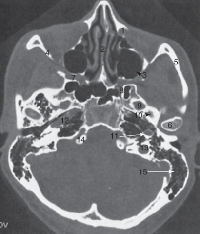
Free
(Multiple Choice)
4.8/5 (35)
Correct Answer:Verified
B
In children, a steeply angled plane is used to scan the brain to avoid radiating the ocular lens.
Free
(True/False)
4.8/5 (42)
Correct Answer:Verified
True
Use the following figure to answer the questions:
-The falx cerebri is labeled: 
(Multiple Choice)
4.8/5 (36)
Spine imaging is acquired:
I. by tilting the gantry
II. perpendicular to the tabletop
III. with the patient in a lateral position
(Multiple Choice)
4.9/5 (42)
Major indications for computed tomography imaging of the head include: I. trauma
II. stroke
III. metastatic disease
(Multiple Choice)
4.9/5 (28)
Use the following figure to answer the questions:
-The anterior (frontal) horn of the lateral ventricle is labeled as: 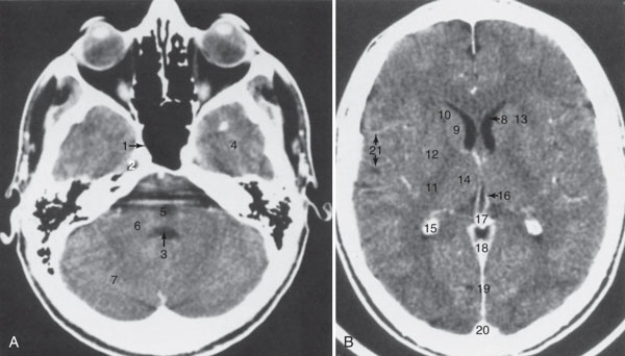
(Multiple Choice)
4.9/5 (32)
All of the following are algorithms used for the brain, neck, and spine except:
(Multiple Choice)
4.9/5 (38)
Use the following figure to answer the questions:
-The sphenoid sinus is labeled: 
(Multiple Choice)
5.0/5 (41)
Which of the following is the most common condition evaluated by repeated computed tomographic head scanning?
(Multiple Choice)
4.8/5 (32)
Use the following figure to answer the questions:
-The common carotid artery is labeled: 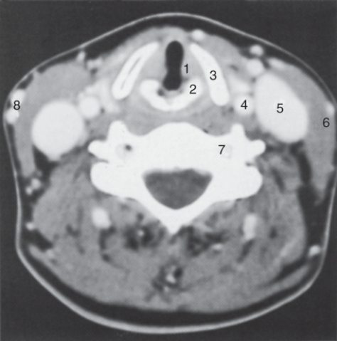
(Multiple Choice)
4.8/5 (36)
A standard dose of contrast is administered for each type of examination on the basis of the patient's weight, up to a maximum.
(True/False)
4.9/5 (38)
Use the following figures to answer the questions:
-The intervertebral disk is labeled: 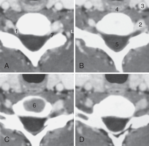
(Multiple Choice)
4.9/5 (40)
Use the following figure to answer the questions:
-The structure labeled as 1 is the: 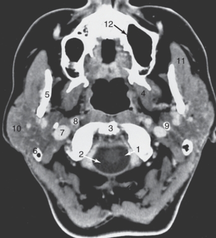
(Multiple Choice)
4.9/5 (27)
Asnwer the Questions according to the figure below:
-The lateral orbital wall is labeled: 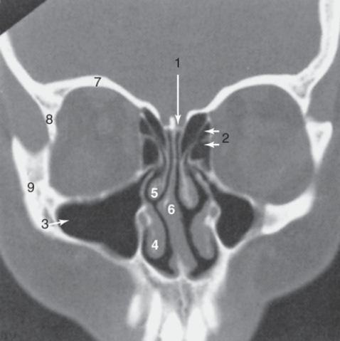
(Multiple Choice)
4.7/5 (41)
Contrast enhancement is well visualized on bone algorithm or bone windows.
(True/False)
4.9/5 (23)
Use the following figure to answer the questions:
-The thalamus is labeled: 
(Multiple Choice)
4.9/5 (31)
Use the following figure to answer the questions:
-The parotid gland is labeled: 
(Multiple Choice)
4.9/5 (35)
Computed tomography provides rapid information about traumatic injuries including:
I. contusions
II. fractures
III. hematomas
(Multiple Choice)
4.9/5 (42)
Filters
- Essay(0)
- Multiple Choice(0)
- Short Answer(0)
- True False(0)
- Matching(0)