Short Answer
To study chromosome movement during anaphase in mammals, you have injected fluorescently labeled tubulin subunits into cells such that microtubules can be seen under a fluorescence microscope. You then use a relatively strong laser beam to bleach the fluorescent dyes in a limited area of a metaphase cell, as indicated in the schematic diagram below, and follow the progression of mitosis under the microscope by time-lapse imaging. You then use the images to measure the change in distance between various components, and plot the results in the graph below. Answer the following question(s) based on these results.
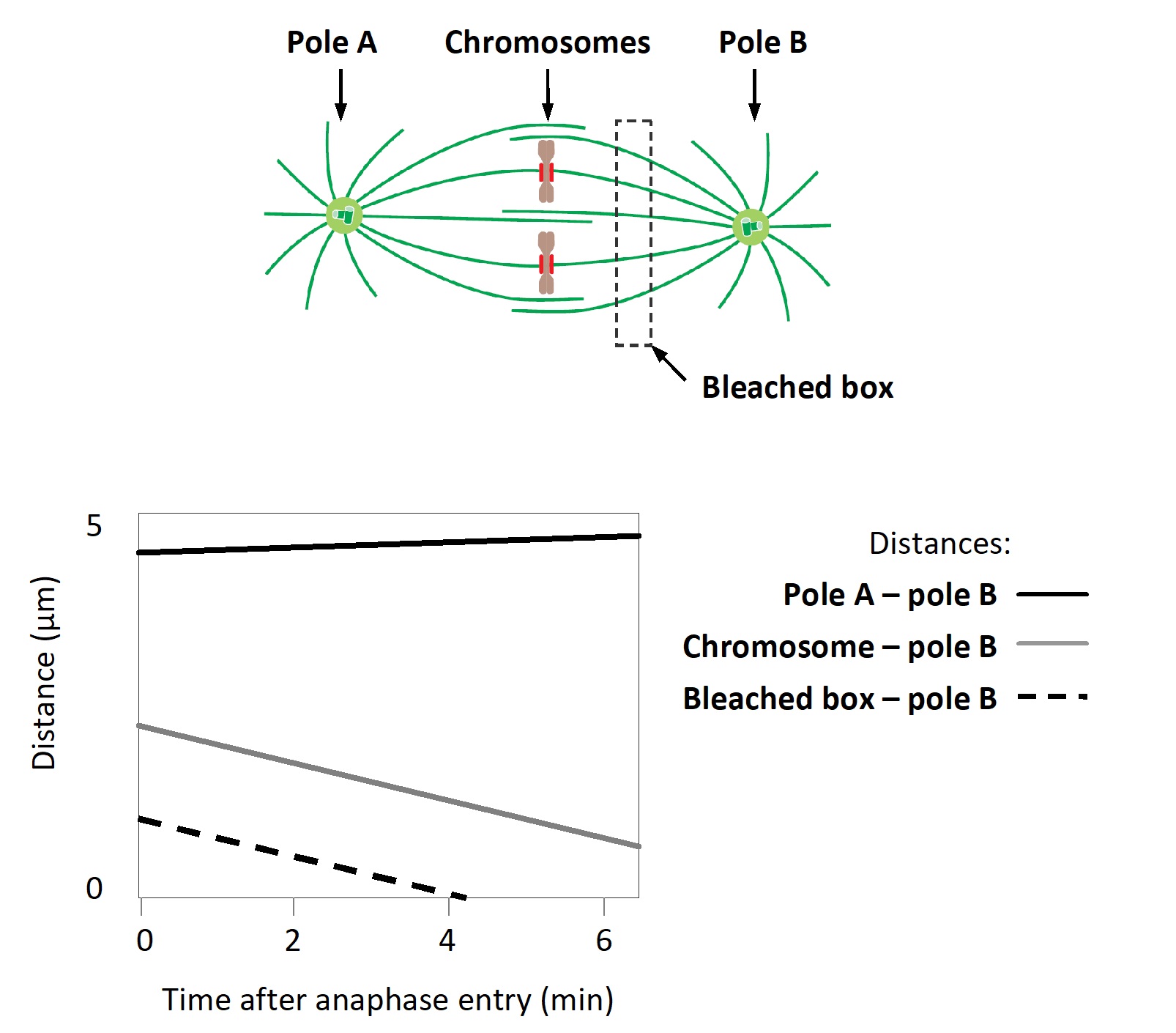
-Based on the results from your experiment, which force is dominant in chromosome movement in this cell: polar ejection force (E), microtubule flux (F), or kinetochore microtubule plus-end depolymerization (P)? Write down E, F, or P as your answer.
Correct Answer:

Verified
In these cells, the chromosomes seem to...View Answer
Unlock this answer now
Get Access to more Verified Answers free of charge
Correct Answer:
Verified
View Answer
Unlock this answer now
Get Access to more Verified Answers free of charge
Q11: Which one better supports cell proliferation when
Q12: To study chromosome movement during anaphase in
Q13: Three models for contractile-ring positioning in animal
Q14: How is Cdc20-APC/C similar to Cdh1-APC/C?<br>A) They
Q15: Which of the following cell populations in
Q17: Formin nucleates the growth of parallel actin
Q18: Consider two kinesin motor proteins at the
Q19: Mammalian Cdk inhibitor proteins (CKIs) can be
Q20: You have been studying the effect of
Q21: Three models for contractile-ring positioning in animal