Multiple Choice
What projection (method) is demonstrated in the image below used to evaluate the cranium?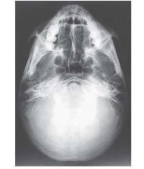
A) PA axial (Caldwell)
B) AP axial (Towne)
C) PA axial (Haas)
D) SMV (Shüller)
Correct Answer:

Verified
Correct Answer:
Verified
Related Questions
Q30: What type of joint is the TMJ?<br>A)
Q33: The part of the cranial base identified
Q34: The axiolateral oblique projection is used to
Q36: Which two facial bones form the roof
Q37: The suture identified on the figure below
Q40: What projection and anatomy is demonstrated in
Q51: Which line should be placed parallel to
Q89: The largest sinus is the:<br>A) frontal.<br>B) maxillary.<br>C)
Q114: The midsagittal plane of the head is
Q133: Which of the following is placed perpendicular