Multiple Choice
What projection and anatomy is demonstrated in the image below?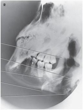
A) AP axial of the TMJs
B) PA axial of the mandibular rami
C) PA of the mandibular body
D) Axiolateral oblique of the mandibular body
Correct Answer:

Verified
Correct Answer:
Verified
Related Questions
Q35: What projection (method) is demonstrated in the
Q36: Which two facial bones form the roof
Q37: The suture identified on the figure below
Q42: Which projections will demonstrate the ethmoidal sinuses?<br>1)Lateral<br>2)PA
Q44: Which line is placed perpendicular to the
Q45: The letter A in the image below
Q77: For a lateral projection of the facial
Q114: The midsagittal plane of the head is
Q129: At which level will the central ray
Q133: Which of the following is placed perpendicular