Multiple Choice
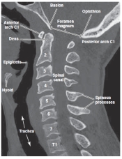
-Refer to the figure. The spinal canal is visible in its full anteroposterior diameter. Why is this possible?
A) This slice is reformatted at the midline
B) Bony pedicles and lamina are not well visualized on CT
C) The spinal cord has a greater density than the spinal canal
D) The spinal canal is only visible on an axial view.
Correct Answer:

Verified
Correct Answer:
Verified
Q2: <img src="https://d2lvgg3v3hfg70.cloudfront.net/TB11028/.jpg" alt=" -Refer to the
Q3: <img src="https://d2lvgg3v3hfg70.cloudfront.net/TB11028/.jpg" alt=" -Refer to the
Q4: The hallmarks of degenerative joint disease in
Q5: What radiographic view is used to screen
Q6: <img src="https://d2lvgg3v3hfg70.cloudfront.net/TB11028/.jpg" alt=" -Refer to the
Q7: Degenerative disk disease is seen on the
Q8: <img src="https://d2lvgg3v3hfg70.cloudfront.net/TB11028/.jpg" alt=" -Refer to the
Q9: <img src="https://d2lvgg3v3hfg70.cloudfront.net/TB11028/.jpg" alt=" -Refer to the
Q10: Foraminal encroachment in the cervical spine would