Essay
In the 1980s, a mutant strain of mice was identified, carrying amino acid changes in the MHC class II gene. This mutant strain was derived from C57Bl/6 mice, which carry the H-2b haplotype. Inbred H-2b mice express only one MHC class II protein, called Ab. The mutant strain, called 'bm12' was found to have 3 amino acid changes in the Ab protein, at positions 67, 70, and 71 of the Aβ chain. The positions of these amino acid changes on the MHC class II structure are shown below by the red circles in Figure . On the right, the side view diagram of MHC class II shows the direction of these three amino acid side chains. 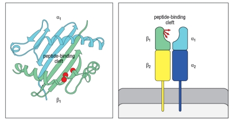
Initial experiments with wild-type C57Bl/6 mice and bm12 mice showed that the wild-type mice made a robust CD4 T cell response after immunization with the insulin protein isolated from a cow; in contrast, the bm12 mice failed to make any detectable response to this foreign protein. Epitope mapping studies identified amino acid residues 1–14 of the bovine insulin A chain as the peptide recognized by CD4 T cells from wild-type mice.
a) What is the most likely explanation for the failure of bm12 mice to make a CD4 T cell response to bovine insulin?
In a second set of experiments, T cells from wild-type (WT) or bm12 mice were mixed in vitro with antigen-presenting cells (APCs), in the presence or absence of the superantigen staphylococcal enterotoxin B (SEB), and T cell proliferation was measured. The data from these experiments are shown in Figure. 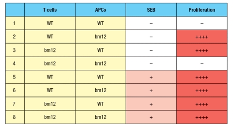
b) What is the explanation for the results in Rows 1–4 of the table?
c) Why does the T cell response to SEB (Rows 5–8) show a different pattern than the response to bovine insulin?
d) In the table above, T cell proliferation was measured after 4 days of incubation of T cells, APCs, +/- SEB. If one isolated the T cells at the end of the incubation for the six conditions in which robust proliferation was seen (Rows 2, 3, 5-8), and stained the T cells with each antibody (separately) from a panel of antibodies that recognize each of the mouse V domains (i.e., an antibody to V 1, an antibody to V 2, etc), what result would be expected?
Correct Answer:

Verified
a) The bovine insulin A (1-14) peptide m...View Answer
Unlock this answer now
Get Access to more Verified Answers free of charge
Correct Answer:
Verified
View Answer
Unlock this answer now
Get Access to more Verified Answers free of charge
Q20: Three major cell types, dendritic cells, macrophages,
Q21: The adaptive immune system uses multiple strategies
Q22: Several types of pathogens encode proteins that
Q23: The extensive polymorphism of MHC genes in
Q24: Alloreactivity refers to the ability of T
Q25: MHC polymorphism at individual MHC genes appears
Q26: The MHC locus encodes a large number
Q28: Multiple mechanisms contribute to create a wide
Q29: The MARCH-1 E3-ubiquitin ligase is expressed in
Q30: The diagram in Figure shows a