Exam 17: The Cardiovascular System I: the Heart
Exam 1: Introduction to Anatomy Physiology101 Questions
Exam 2: The Chemistry of Life117 Questions
Exam 3: The Cell117 Questions
Exam 4: Histology98 Questions
Exam 5: The Integumentary System103 Questions
Exam 6: Bones and Bone Tissue96 Questions
Exam 7: The Skeletal System121 Questions
Exam 8: Articulations112 Questions
Exam 9: The Muscular System118 Questions
Exam 10: Muscle Tissue Physiology104 Questions
Exam 11: Introduction to the Nervous System Nervous Tissue109 Questions
Exam 12: The Central Nervous System125 Questions
Exam 13: The Peripheral Nervous System113 Questions
Exam 14: The Autonomic Nervous System Homeostasis91 Questions
Exam 15: The Special Senses127 Questions
Exam 16: The Endocrine System130 Questions
Exam 17: The Cardiovascular System I: the Heart128 Questions
Exam 18: The Cardiovascular System Ii: Blood Vessels120 Questions
Exam 19: Blood103 Questions
Exam 20: The Lymphatic System Immunity125 Questions
Exam 21: The Respiratory System110 Questions
Exam 22: The Digestive System115 Questions
Exam 23: Metabolism Nutrition116 Questions
Exam 24: The Urinary System115 Questions
Exam 25: Fluid, Electrolyte Acid-Base Homeostasis105 Questions
Exam 26: The Reproductive System120 Questions
Exam 27: Development Heredity118 Questions
Select questions type
Match the following with the waves, segments, or intervals on the electrocardiogram (ECG).
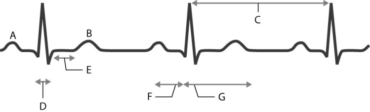 -Identify the P -R interval.
(Short Answer)
4.7/5  (42)
(42)
The P wave on an electrocardiogram (ECG) represents the depolarization of cells in the:
(Multiple Choice)
5.0/5 (48)
Which wave on the electrocardiogram (ECG) represents ventricular depolarization?
(Multiple Choice)
4.7/5 (44)
Describe the pathway of the action potential through the cardiac conduction system.
(Essay)
4.8/5 (27)
Calculate cardiac output if the heart rate is 85 beats/minute, end -diastolic volume (EDV) is 130 ml, and end -systolic volume (ESV) is 60 ml.
(Multiple Choice)
4.9/5 (40)
Stroke volume (SV) can be calculated by subtracting the end -diastolic volume (EDV) from the end -systolic volume (ESV).
(True/False)
4.8/5 (36)
Calculate cardiac output given an end -diastolic volume of 140 ml, an end -systolic volume of 60 ml, and a heart rate of 85 beats/minute.
(Essay)
4.8/5 (39)
There are two phases of the cardiac cycle in which all four heart valves are open: isovolumetric contraction phase and the isovolumetric relaxation phase.
(True/False)
4.7/5 (40)
Trace the pathway of blood flow through the heart. Begin and end the pathway with the systemic capillaries.
(Essay)
4.8/5 (40)
Explain the roles of the right side and left side of the heart as two separate pumps.
(Essay)
4.9/5 (41)
Match the following structures with the internal heart anatomy.
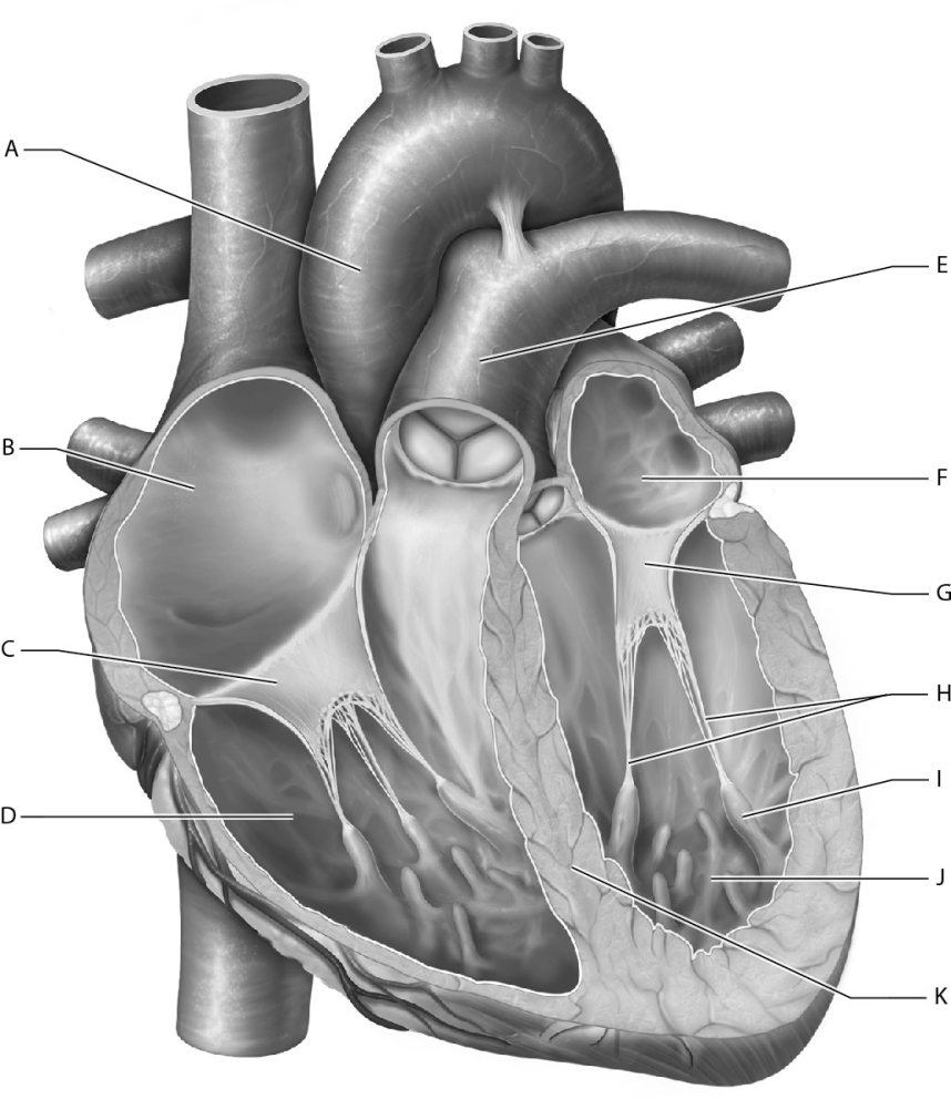 -Identify the interventricular septum.
(Short Answer)
4.8/5 (33)
Explain how contractile cells and pacemaker cells differ in cardiac muscle tissue.
(Essay)
4.7/5 (36)
What characteristic differentiates cardiac muscle cells from skeletal muscle cells?
(Multiple Choice)
4.8/5 (46)
Match the following with the waves, segments, or intervals on the electrocardiogram (ECG).
-Identify the S -T segment.
(Short Answer)
4.8/5 (31)
Match the following with the waves, segments, or intervals on the electrocardiogram (ECG).
-Identify the wave representing depolarization of all cells within the atria except the SA node.
(Short Answer)
4.8/5 (40)
What surface groove separates the right and left ventricles?
(Multiple Choice)
4.8/5 (39)
During what part of the action potential will calcium ions enter the contractile cell?
(Multiple Choice)
4.8/5 (38)
Filters
- Essay(0)
- Multiple Choice(0)
- Short Answer(0)
- True False(0)
- Matching(0)