Exam 2: The First Steps in Vision: From Light to Neural Signals
People with _______ do not require an optical correction to see normally.
D
When light strikes a photoreceptor, what happens to its electrical potential?
A
Describe the transmission of information in the retina from photoreceptors to the optic nerve. What cells are functioning in what order, and how do they transform visual information on the way to the brain?
Once light strikes the outer segment of photoreceptors in the retina, a chromophore captures the light molecule which begins the process of photoactivation, causing the photoreceptor to become hyperpolarized. The graded potentials of the photoreceptor that result from photoactivation are sent on to the bipolar cells, which may be either diffuse bipolar cells or midget bipolar cells. Diffuse bipolar cells connect to multiple cones while midget bipolar cells connect to only a single cone. Horizontal cells connect to the synapse of photoreceptors and bipolar cells and help to create the center-surround receptive field organization through lateral inhibition. ON bipolar cells respond to increases in light and OFF bipolar cells respond to decreases in light. Bipolar cells connect to P and M ganglion cells as well as amacrine cells. Amacrine cells modulate the signals coming from bipolar cells to emphasize contrast and improve temporal processing. P ganglion cells receive excitatory input from midget bipolar cells and feed the parvocellular layer of the lateral geniculate nucleus (LGN). M ganglion cells receive excitatory input from diffuse bipolar cells and feed the magnocellular layer of the LGN Ganglion cells converge at the optic disk where they leave the eye and form the optic nerve. The optic nerve transmits signals from the retina to the LGN.
The dark, circular opening at the center of the eye, where light enters the eye, is called the
Retinal cells called _______ leave the eye via the optic nerve and transmit information to the brain and midbrain.
With regard to retinitis pigmentosa (RP) and age-related macular degeneration (AMD), which would have the greatest impact on photopic (daytime) vision, and why?
Which of the following is a unit of measurement of the optic power of a lens?
If the optic nerve is severed, which retinal cells are damaged?
Refer to the figure.
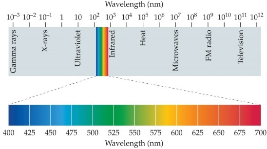 The bottom part of the figure shows the
Which stimulus would optimally activate an OFF-center ganglion cell?
The transparent "window" on the outer part of the eye that allows light into the eyeball is called the
The high-resolution part of the eye that is used for detailed vision is called the
We all have a blind spot in each eye. In normal circumstances, why is it that we do not experience large black empty regions in our visual field?
What are age-related macular degeneration (AMD) and retinitis pigmentosa (RP)? How are they similar and how are they different?
Refer to the figure.
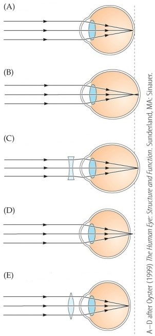 Which part of the figure depicts hyperopia without correction?
With regard to retinitis pigmentosa (RP) and age-related macular degeneration (AMD), which would have the greatest impact on scotopic (nighttime) vision, and why?
Filters
- Essay(0)
- Multiple Choice(0)
- Short Answer(0)
- True False(0)
- Matching(0)