Multiple Choice
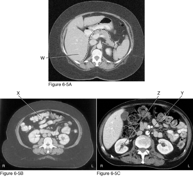
-Figure 6-5A, B, and C are images from an __________.
A) Axial MRI of the abdomen
B) Axial computed tomography scan of the abdomen
C) Axial computed tomography scan of the brain
D) Axial computed tomography scan of the chest
Correct Answer:

Verified
Correct Answer:
Verified
Q2: <img src="https://d2lvgg3v3hfg70.cloudfront.net/TB4823/.jpg" alt=" -The structure indicated
Q3: <img src="https://d2lvgg3v3hfg70.cloudfront.net/TB4823/.jpg" alt=" -What type of
Q4: <img src="https://d2lvgg3v3hfg70.cloudfront.net/TB4823/.jpg" alt=" -The structure indicated
Q5: <img src="https://d2lvgg3v3hfg70.cloudfront.net/TB4823/.jpg" alt=" -The structure marked
Q6: <img src="https://d2lvgg3v3hfg70.cloudfront.net/TB4823/.jpg" alt=" -The structure indicated
Q7: <img src="https://d2lvgg3v3hfg70.cloudfront.net/TB4823/.jpg" alt=" -The structure indicated
Q8: <img src="https://d2lvgg3v3hfg70.cloudfront.net/TB4823/.jpg" alt=" -The structure indicated
Q9: <img src="https://d2lvgg3v3hfg70.cloudfront.net/TB4823/.jpg" alt=" -Identify the diagnostic
Q10: <img src="https://d2lvgg3v3hfg70.cloudfront.net/TB4823/.jpg" alt=" -What type of
Q11: <img src="https://d2lvgg3v3hfg70.cloudfront.net/TB4823/.jpg" alt=" -The structure indicated