Exam 9: Visualizing Cells
Exam 1: Cells and Genomes34 Questions
Exam 2: Cell Chemistry and Bioenergetics54 Questions
Exam 3: Proteins52 Questions
Exam 4: DNA, Chromosomes, and Genomes57 Questions
Exam 5: DNA Replication, Repair, and Recombination51 Questions
Exam 6: How Cells Read the Genome: From DNA to Protein58 Questions
Exam 7: Control of Gene Expression62 Questions
Exam 8: Analyzing Cells, Molecules, and Systems95 Questions
Exam 9: Visualizing Cells29 Questions
Exam 10: Membrane Structure26 Questions
Exam 11: Membrane Transport of Small Molecules and the Electrical Properties of Membranes46 Questions
Exam 12: Intracellular Compartments and Protein Sorting46 Questions
Exam 13: Intracellular Membrane Traffic54 Questions
Exam 14: Energy Conversion: Mitochondria and Chloroplasts49 Questions
Exam 15: Cell Signaling63 Questions
Exam 16: The Cytoskeleton75 Questions
Exam 17: The Cell Cycle57 Questions
Exam 18: Cell Death12 Questions
Exam 19: Cell Junctions and the Extracellular Matrix56 Questions
Exam 20: Cancer50 Questions
Exam 21: Development of Multicellular Organisms61 Questions
Exam 22: Stem Cells and Tissue Renewal45 Questions
Exam 23: Pathogens and Infection32 Questions
Exam 24: The Innate and Adaptive Immune Systems47 Questions
Select questions type
Atomic force microscopy (AFM) is used in an experiment to unfold a multidomain protein by applying mechanical force. The protein contains several copies of an immunoglobulin domain that are unfolded one by one as the two ends of the molecule (one attached to a cover slip, and the other to the AFM tip) are being pulled apart, resulting in the "sawtooth" force-extension curves shown below. The same experiment is done twice, once in the presence and once in the absence of a chaperone protein that stabilizes the immunoglobulin domains. Answer the following questions based on this graph.
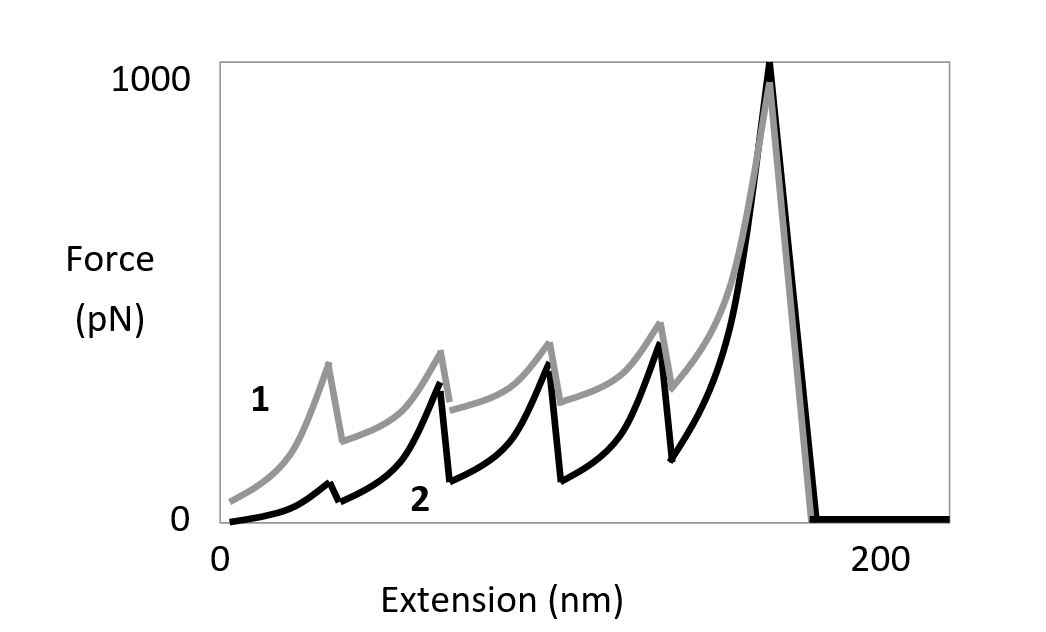 -According to the force-extension graph, which curve (1 or 2) would you expect to correspond to the reaction in the presence of the chaperone protein? Write down 1 or 2 as your answer.
Free
(Short Answer)
4.8/5  (41)
(41)
Correct Answer: Verified
1
When the gene encoding a certain cytoskeleton protein is deleted, the resulting mutant cells round up and do not form their normal appendages. These mutants can be rescued when a gene encoding an N-terminal green fluorescent protein (GFP) fusion of the protein is expressed, but not when a gene encoding a C-terminal GFP fusion is expressed. Which fusion protein (N or C) is appropriate to use in studying cellular localization and activity? Write down N or C as your answer.
Free
(Short Answer)
4.8/5 (34)
Correct Answer:Verified
N
Atomic force microscopy (AFM) is used in an experiment to unfold a multidomain protein by applying mechanical force. The protein contains several copies of an immunoglobulin domain that are unfolded one by one as the two ends of the molecule (one attached to a cover slip, and the other to the AFM tip) are being pulled apart, resulting in the "sawtooth" force-extension curves shown below. The same experiment is done twice, once in the presence and once in the absence of a chaperone protein that stabilizes the immunoglobulin domains. Answer the following questions based on this graph.
-According to the force-extension graph, how many immunoglobulin domains are unfolded in each of these experiments? Write down the number as your answer, e.g. 9.
Free
(Short Answer)
4.8/5 (32)
Correct Answer:Verified
4
What is the advantage of using quantum dots as an alternative to organic fluorochromes such as Cy3 and Alexa dyes?
(Multiple Choice)
4.8/5 (34)
In the diagram below, a logarithmic scale of sizes is shown. Indicate which of the sizes indicated (A to H) better corresponds to the dimensions of each of the following. Your answer would be a three-letter string composed of letters A to H only, e.g. HCG.
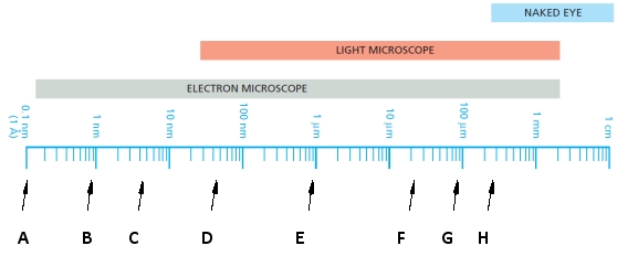 ( ) A bacterium
( ) An animal cell
( ) A globular protein
( ) A bacterium
( ) An animal cell
( ) A globular protein
(Short Answer)
4.8/5 (39)
Electron microscopy samples are often chemically fixed before dehydration, resin-embedding, and sectioning. But they can also be "fixed" by rapid freezing, in a way that precludes ice-crystal formation, to ensure minimal damage to the original cell structures. How can this be done?
(Multiple Choice)
4.7/5 (27)
Indicate whether each of the following descriptions better applies to the scanning (S) or transmission (T) electron microscopy techniques. Your answer would be a four-letter string composed of letters S and T only, e.g. TSTS.
( ) It generally has a greater depth of field.
( ) It is usually smaller, cheaper, and simpler.
( ) It detects electrons that are scattered or emitted from the specimen.
( ) It is used to create electron-microscope tomograms.
(Short Answer)
4.9/5 (28)
Indicate true (T) and false (F) statements below regarding electron microscopy. Your answer would be a four-letter string composed of letters T and F only, e.g. FFFF.
( ) Depending on acceleration voltage, the resolution limit of an electron microscope can be as small as 0.05 nm.
( ) The emission gun and the magnetic coils in an electron microscope are analogous to the light source and the glass lenses in a light microscope, respectively.
( ) Contrast in specimens for electron microscopy can be achieved using electron-dense material.
( ) For biological samples, the effective resolution of electron microscopy is about 1 nm.
(Short Answer)
5.0/5 (31)
You have generated strains of Drosophila melanogaster that are expected to show interesting developmental phenotypes such as misplaced organs in the adult fly. However, some of these phenotypes are not readily seen with light microscopy. You therefore fix each mutant fly, dry it, coat it with a thin layer of gold, and place the entire fly into an electron microscope for viewing. What type of microscope are you using? Write down SEM or TEM as your answer.
(Short Answer)
4.9/5 (40)
The presence of which of the following provides the sample with the lowest electron density?
(Multiple Choice)
4.8/5 (29)
Consider an engineered chimeric protein made from fusion of three proteins: a blue fluorescent protein (BFP), a calmodulin-binding peptide, and a green fluorescent protein (GFP). Calmodulin is an abundant calcium-binding protein in eukaryotes. Once bound to calcium ions, it can recognize the calmodulin-binding peptide in the fusion protein, change conformation, wrap around the peptide, and bring the BFP and GFP components in close proximity. This results in fluorescence resonance energy transfer (FRET) between BFP and GFP. Accordingly, the fusion protein …
(Multiple Choice)
4.8/5 (36)
Indicate whether each of the following descriptions better applies to SIM (S), STED (T), or STORM/PALM (P) superresolution techniques. Your answer would be a four-letter string composed of letters S, T, and P only, e.g. PPTS.
( ) It switches on and off individual fluorophores at random over time to accurately determine their position.
( ) It creates a moiré pattern from the interference of the illuminating pattern and the sample features.
( ) It doubles the resolution of conventional fluorescence microscopy.
( ) It limits excitation to the fluorophores that are located at the center of the focal point by using a doughnut-shaped beam in addition to the excitation beam.
(Short Answer)
4.9/5 (34)
Which microscopy set-up uses a longer wavelength of light than usually excites a particular fluorophore? Which one allows researchers to peek deeper into biological samples?
(Multiple Choice)
4.7/5 (28)
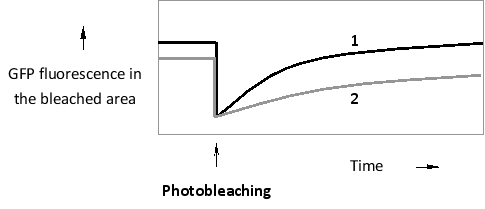
A certain GTP-binding protein can exist in two main states. When bound to GDP, it is mostly cytosolic. In its GTP-bound form, however, it associates with the cytosolic face of the endoplasmic reticulum (ER) membrane, where it hydrolyzes the bound GTP after a short delay and is released again into the cytosol. You have created and expressed green fluorescent protein (GFP) fusions of the wild-type protein, as well as that of a mutant protein that does not bind GTP as readily as the wild type. You then perform a fluorescence recovery after photobleaching (FRAP) experiment by photobleaching a small area of the ER membrane and measuring GFP fluorescence recovery over time. According to the results below, which curve (1 or 2) do you think corresponds to the wild-type fusion protein? Write down 1 or 2 as your answer.
(Short Answer)
4.9/5 (32)
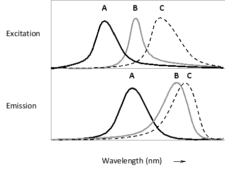
Given the absorption and emission spectra of three fluorescent dyes in the simplified diagrams below, which pair of dyes is better suited for a fluorescence resonance energy transfer (FRET) study? Write down AB, BC, or AC as your answer.
(Short Answer)
4.9/5 (40)
Indicate true (T) and false (F) statements below regarding light and light microscopy. Your answer would be a four-letter string composed of letters T and F only, e.g. FFFF.
( ) Two light waves of the same amplitude and frequency will completely cancel each other out if not perfectly in phase.
( ) If the refractive index of a medium is 1.1, light travels in a vacuum 1.1 times faster than it does in the medium.
( ) The limit of resolution for conventional light microscopy is approximately 0.4 µm, corresponding to the wavelength of violet light.
( ) A light-emitting particle can be detected with a light microscope even if it is several times smaller than the resolution limit of the microscope.
(Short Answer)
4.9/5 (38)
Single-molecule detection by fluorescence microscopy is limited by the presence of an excess of out-of-focus fluorescent molecules. How does a TIRF microscope uniquely overcome this limitation?
(Multiple Choice)
4.8/5 (26)
Two approaches have been devised to deal with the problem of blurring in light microscopy with thicker samples. Indicate whether each of the following descriptions better applies to confocal design (C) or image deconvolution (D). Your answer would be a three-letter string composed of letters C and D only, e.g. CCD.
( ) It is normally faster.
( ) It requires a higher degree of sample illumination.
( ) It can be used to obtain images from relatively deeper parts of the specimen.
(Short Answer)
4.8/5 (27)
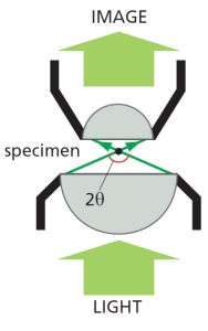
The following schematic diagram shows the path of light rays passing through in a light microscope. If the angular width of the cone of rays collected by the objective lens (2?) is increased, would the resolution improve (I; i.e. the resolution limit decreases) or deteriorate (D)? Write down I or D as your answer.
(Short Answer)
4.8/5 (26)
Indicate whether you would use a fluorescent organic molecule (O), in situ hybridization (H), or a coupled fluorescent protein (P) to visualize the cells and their molecules in each of the following cases. Your answer would be a five-letter string composed of letters O, H, and P only, e.g. OHOOO.
( ) You would like to see where in the early Drosophila embryo the mRNA encoding a certain transcription regulator is located.
( ) You would like to see the nuclei and count them in an early mouse embryo.
( ) You would like to visualize chromosome 3 in a human cell culture derived from a patient's tissue, based on specific sequences present on this chromosome.
( ) You would like to observe the oscillations in Ca²? ions inside a fertilized frog egg.
( ) You would like to compare the localization of two transcription regulatory proteins in cultured human T cells.
(Short Answer)
4.8/5 (40)
Filters
- Essay(0)
- Multiple Choice(0)
- Short Answer(0)
- True False(0)
- Matching(0)