Multiple Choice
In an unfolded (random coil) protein, amino acid residues are exposed to the solvent and share more or less the same environment, whereas each residue in a folded protein has its own unique neighborhood. This fact can be exploited in using NMR to study protein folding. The two schematic diagrams below represent two-dimensional NMR spectra for the same protein in either its folded (native) or unfolded state. The chemical shifts, which depend on the local neighborhood of each atom, are plotted in these diagrams. Which diagram (A or B) do you think corresponds to the folded protein?
A) 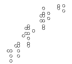
B) 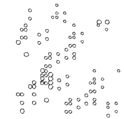
Correct Answer:

Verified
Correct Answer:
Verified
Q85: A piece of DNA has been sequenced
Q86: Sort the following cellular components to reflect
Q87: In purifying proteins by column chromatography, elution
Q88: RNA-seq and ribosome profiling experiments have been
Q89: You have grown cultures of the yeast
Q91: Some feed-forward motifs are capable of generating
Q92: There are six possible reading frames for
Q93: In the following schematic graph, the fraction
Q94: A protein made from an expression vector
Q95: The following graph shows the change in