Exam 2: Observing the Microbial Cell
Exam 1: Microbial Life: Origin and Discovery69 Questions
Exam 2: Observing the Microbial Cell69 Questions
Exam 3: Cell Structure and Function72 Questions
Exam 4: Bacterial Culture, Growth, and Development70 Questions
Exam 5: Environmental Influences and Control of Microbial Growth70 Questions
Exam 6: Viruses70 Questions
Exam 7: Genomes and Chromosomes70 Questions
Exam 8: Transcription, Translation, and Bioinformatics76 Questions
Exam 9: Gene Transfer, Mutations, and Genome Evolution72 Questions
Exam 10: Molecular Regulation73 Questions
Exam 11: Viral Molecular Biology70 Questions
Exam 12: Biotechniques and Synthetic Biology72 Questions
Exam 13: Energetics and Catabolism77 Questions
Exam 14: Electron Flow in Organotrophy, Lithotrophy, and Phototrophy70 Questions
Exam 15: Biosynthesis70 Questions
Exam 16: Food and Industrial Microbiology73 Questions
Exam 17: Origins and Evolution70 Questions
Exam 18: Bacterial Diversity71 Questions
Exam 19: Archaeal Diversity70 Questions
Exam 20: Eukaryotic Diversity70 Questions
Exam 21: Microbial Ecology70 Questions
Exam 22: Microbes in Global Elemental Cycles70 Questions
Exam 23: Human Microbiota and Innate Immunity70 Questions
Exam 24: The Adaptive Immune Response70 Questions
Exam 25: Microbial Pathogenesis70 Questions
Exam 26: Microbial Diseases69 Questions
Exam 27: Antimicrobial Therapy72 Questions
Exam 28: Clinical Microbiology and Epidemiology75 Questions
Select questions type
Which of the following techniques can visualize bacteria without focusing electromagnetic radiation?
Free
(Multiple Choice)
4.8/5  (34)
(34)
Correct Answer: Verified
D
Why do some bacteria appear purple after being Gram stained, while others appear pink?
Free
(Essay)
4.7/5 (46)
Correct Answer:Verified
Gram-negative cells have a few layers of peptidoglycan cell wall and an outer lipopolysaccharide membrane. Gram-positive organisms have several layers of peptidoglycan and no outer membrane. The multiple layers of peptidoglycan retain the crystal violet-iodine complex, so appear purple. Gram-negative cells do not retain the crystal violet because there are few layers of peptidoglycan and the outer membrane is disrupted by the decolorizer.
If your eyes had photoreceptors packed as closely as an eagle's (about eight times greater than humans), would you be able to resolve a virus (100 nm in size) using a light microscope? Why or why not?
Free
(Essay)
4.9/5 (38)
Correct Answer:Verified
No. Although your resolving power would be much improved, the light microscope's power will still be limited by the wavelengths of light that you can see (roughly 400 nm for human eyes). Objects less than 400 nm cannot be resolved by light in the visible spectrum.
Which of the following is best visualized using a negative stain?
(Multiple Choice)
4.9/5 (39)
Which of these techniques would provide the best resolution of an enzyme's structure?
(Multiple Choice)
4.8/5 (37)
Fluorescence microscopy using labeled antibodies is referred to as
(Multiple Choice)
4.7/5 (38)
Which type of microscopy is used to view the internal structures of a specimen?
(Multiple Choice)
4.7/5 (39)
The part of the human eye that is MOST involved in resolving an image is the
(Multiple Choice)
4.9/5 (38)
When two waves are out of phase by ________ wavelength, they produce destructive interference, canceling each other's amplitude and resulting in contrast in the image.
(Multiple Choice)
4.9/5 (33)
Which of these series arranges microbes from smallest to largest?
(Multiple Choice)
4.7/5 (38)
The spots recorded on film during X-ray diffraction analyses are due to
(Multiple Choice)
4.9/5 (38)
What is the best explanation for a Gram-positive bacterium appearing pink after performing a Gram stain?
(Multiple Choice)
5.0/5 (41)
Which of the following methods is used to generate the structure shown below? 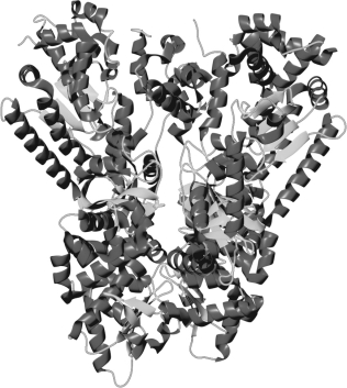
(Multiple Choice)
4.8/5 (32)
Microbes were detected long before the invention of the microscope. How could this be?
(Essay)
4.7/5 (42)
Which type of microscopy is used to identify the 3-D structure of biofilms?
(Multiple Choice)
4.9/5 (33)
Describe three methods of sample preparation for electron microscopy. Which method would cause the fewest artifacts? Why?
(Essay)
4.9/5 (34)
In which type of microscopy do dust particles interfere the MOST?
(Multiple Choice)
4.9/5 (36)
Filters
- Essay(0)
- Multiple Choice(0)
- Short Answer(0)
- True False(0)
- Matching(0)