Exam 2: Observing the Microbial Cell
Exam 1: Microbial Life: Origin and Discovery69 Questions
Exam 2: Observing the Microbial Cell69 Questions
Exam 3: Cell Structure and Function72 Questions
Exam 4: Bacterial Culture, Growth, and Development70 Questions
Exam 5: Environmental Influences and Control of Microbial Growth70 Questions
Exam 6: Viruses70 Questions
Exam 7: Genomes and Chromosomes70 Questions
Exam 8: Transcription, Translation, and Bioinformatics76 Questions
Exam 9: Gene Transfer, Mutations, and Genome Evolution72 Questions
Exam 10: Molecular Regulation73 Questions
Exam 11: Viral Molecular Biology70 Questions
Exam 12: Biotechniques and Synthetic Biology72 Questions
Exam 13: Energetics and Catabolism77 Questions
Exam 14: Electron Flow in Organotrophy, Lithotrophy, and Phototrophy70 Questions
Exam 15: Biosynthesis70 Questions
Exam 16: Food and Industrial Microbiology73 Questions
Exam 17: Origins and Evolution70 Questions
Exam 18: Bacterial Diversity71 Questions
Exam 19: Archaeal Diversity70 Questions
Exam 20: Eukaryotic Diversity70 Questions
Exam 21: Microbial Ecology70 Questions
Exam 22: Microbes in Global Elemental Cycles70 Questions
Exam 23: Human Microbiota and Innate Immunity70 Questions
Exam 24: The Adaptive Immune Response70 Questions
Exam 25: Microbial Pathogenesis70 Questions
Exam 26: Microbial Diseases69 Questions
Exam 27: Antimicrobial Therapy72 Questions
Exam 28: Clinical Microbiology and Epidemiology75 Questions
Select questions type
A(n) ________ acts to vary the diameter of the light column in a light microscope.
(Multiple Choice)
5.0/5  (42)
(42)
Which type of microscopy is particularly useful to study the surfaces of live bacteria?
(Multiple Choice)
5.0/5 (35)
Most electron micrographs in microbiology textbooks are in color. Is this normal for an electron micrograph? Why or why not?
(Essay)
4.9/5 (36)
Transmission electron microscopy commonly has a resolution of ________ the highest resolution possible for light microscopy.
(Multiple Choice)
4.8/5 (40)
Define a fluorophore and give three examples of how it can be used to label cells.
(Essay)
4.7/5 (35)
What color are Gram-positive and Gram-negative cells when properly Gram stained? For each step of the Gram-stain procedure, predict the colors of a Gram-positive or Gram-negative cell if that step were omitted during staining. Explain your reasoning.
(Essay)
4.9/5 (39)
Compare and contrast a simple stain (like methylene blue) with the Gram stain. What information about a microbial sample can be collected with each?
(Essay)
4.8/5 (38)
Give a few reasons why living organisms may not be observed by transmission electron microscopy (TEM) or scanning electron microscopy (SEM).
(Essay)
4.8/5 (39)
Wavelength interference results in small observed objects (like bacteria) being surrounded by
(Multiple Choice)
4.9/5 (25)
Which two components of the Gram stain form a complex that is retained by Gram-positive cells?
(Multiple Choice)
5.0/5 (35)
Which of the following is an advantage of using chemical imaging microscopy?
(Multiple Choice)
4.9/5 (45)
Using the figure below, explain how the visualization of molecules occurs through X-ray crystallography.
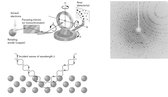
(Essay)
4.7/5 (29)
The knife used to cut embedded specimens for observation by transmission electron microscopy is called a
(Multiple Choice)
4.7/5 (38)
The figure below displays absorption and emission spectra for a fluorophore. Describe the difference between absorption and emission and how this is reflected in the figure.
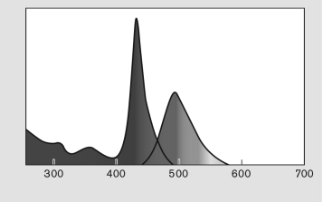
(Essay)
4.9/5 (40)
The figure below shows the Gram-staining process. Describe what is happening at step number 3 relative to bacterial cell walls.
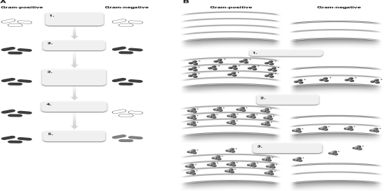
(Essay)
4.9/5 (33)
Compare and contrast the radiation sources, lenses, and image-capturing devices used in light microscopy and transmission electron microscopy.
(Essay)
4.9/5 (39)
Chlamydia trachomatis transmits infection to a new cell via
(Multiple Choice)
4.8/5 (35)
Filters
- Essay(0)
- Multiple Choice(0)
- Short Answer(0)
- True False(0)
- Matching(0)