Exam 4: The Tissue Level of Organization
Exam 1: An Introduction to the Human Body78 Questions
Exam 2: The Chemical Level of Organization81 Questions
Exam 3: The Cellular Level of Organization84 Questions
Exam 4: The Tissue Level of Organization64 Questions
Exam 5: The Integumentary System78 Questions
Exam 6: The Skeletal System: Bone Tissue77 Questions
Exam 7: The Skeletal System: the Axial Skeleton80 Questions
Exam 8: The Skeletal System: The Appendicular Skeleton82 Questions
Exam 9: Joints41 Questions
Exam 10: Muscular Tissue85 Questions
Exam 11: The Muscular System70 Questions
Exam 12: Nervous Tissue17 Questions
Exam 13: The Spinal Cord and Spinal Nerves74 Questions
Exam 14: The Brain and Cranial Nerves72 Questions
Exam 15: The Autonomic Nervous System64 Questions
Exam 16: Sensory, Motor, and Integrative Systems76 Questions
Exam 17: The Special Senses73 Questions
Exam 18: The Endocrine System84 Questions
Exam 19: The Cardiovascular System: the Blood83 Questions
Exam 20: The Cardiovascular System: the Heart82 Questions
Exam 21: The Cardiovascular System: Blood Vessels and Hemodynamics81 Questions
Exam 22: The Lymphatic System and Immunity80 Questions
Exam 23: The Respiratory System85 Questions
Exam 24: The Digestive System85 Questions
Exam 25: Metabolism and Nutrition85 Questions
Exam 26: The Urinary System84 Questions
Exam 27: Fluid, Electrolyte, and Acidbase Homeostasis57 Questions
Exam 28: The Reproductive Systems84 Questions
Exam 29: Development and Inheritance83 Questions
Select questions type
Name and briefly describe the two types of growth seen in cartilage.
Free
(Essay)
4.8/5  (32)
(32)
Correct Answer: Verified
Growth of cartilage can be classified as interstitial or appositional.In interstitial growth,the cartilage increases rapidly in size due to the division of existing chondrocytes and the continuous deposition of increasing amounts of matrix by the chondrocytes.In appositional growth,activity of the cells in the inner chondrogenic layer of the perichondrium leads to growth.Deeper layers divide and cells mature resulting in the matrix accumulating beneath the perichondrium on the outer surface of the cartilage causing it to grow in width.
[dropdown 1]covers and protects surfaces inside and outside of the body.[dropdown 2] help secure most of the epidermal cells together through intermediate filaments,while [dropdown 3]allow communication among the epidermal cells.This tissue will have a [dropdown 4]surface that is at the superficial layer,and a [dropdown 5]surface that is attached to the basement membrane by [dropdown 6].
![[dropdown 1]covers and protects surfaces inside and outside of the body.[dropdown 2] help secure most of the epidermal cells together through intermediate filaments,while [dropdown 3]allow communication among the epidermal cells.This tissue will have a [dropdown 4]surface that is at the superficial layer,and a [dropdown 5]surface that is attached to the basement membrane by [dropdown 6].](https://storage.examlex.com/TB6091/11ee3a7f_b72c_1087_913d_df2d1a47920d_TB6091_11.jpg)
Free
(Short Answer)
4.8/5 (28)
Correct Answer:Verified
1.epithelial
2.desmosomes
3.gap
4.apical
5.basal
6.hemidesmosomes
_______ are abnormal joining of tissues resulting from the formation of scar tissue at a previous site of inflammation or surgical repair.
Free
(Multiple Choice)
4.9/5 (30)
Correct Answer:Verified
A
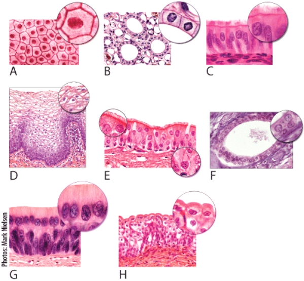
Which of the light micrographs in the following figure shows a type of epithelial tissue whose functions include movement of mucus over their apical surface by ciliary action?
1 B
2 C
3 E

(Multiple Choice)
4.8/5 (39)
Which of the light micrographs shows epithelial tissue that forms thyroid gland and secretes hormones?

(Multiple Choice)
4.8/5 (32)
Which of the light micrographs in the following figure shows the type of epithelial tissue that lines the inside of urinary bladder walls and the outside of the urinary bladder?
(Multiple Choice)
4.8/5 (34)
Match the matrix with the correct connective tissue
Blood: [dropdown 1]
Cartilage: [dropdown 2]
Dense regular: [dropdown 3]
Dropdown choices:
chondroitin sulfate
collagen
plasma
(Short Answer)
4.9/5 (36)
Vitamin C is needed for healthy collagen fibers.What tissue will not be as affected if vitamin C was absent or present in low amounts?
(Multiple Choice)
4.9/5 (35)
The pubic symphysis and intervertebral discs are composed of
(Multiple Choice)
5.0/5 (37)
Which epithelial tissue lines the ducts of sweat glands and oil glands?
(Multiple Choice)
4.8/5 (35)
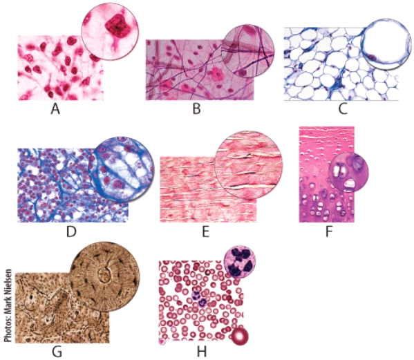
Which figure shows collagen fibers running in a uniformed pattern?
(Multiple Choice)
4.8/5 (37)
Which tissue is found in the stomach? Select all that apply.
(Multiple Choice)
4.7/5 (28)
In the figure shown,light micrograph [___] will be located in red bone marrow and in the spleen to help with filtering.
![In the figure shown,light micrograph [___] will be located in red bone marrow and in the spleen to help with filtering. Dropdown choices: A B C D E F G H I J](https://storage.examlex.com/TB6091/11ea7f55_b8ee_15d0_9ecd_a77877fbbaba_TB6091_00.jpg) Dropdown choices:
A
B
C
D
E
F
G
H
I
J
Dropdown choices:
A
B
C
D
E
F
G
H
I
J
(Short Answer)
4.8/5 (33)
Which of the light micrographs in the following figure shows the type of epithelial tissue whose structure allows it to be stretched or distended?

(Multiple Choice)
4.8/5 (34)
Which cells are considered excitable cells because they are able to produce electrical signals?
(Multiple Choice)
4.8/5 (45)
Filters
- Essay(0)
- Multiple Choice(0)
- Short Answer(0)
- True False(0)
- Matching(0)