Exam 20: The Cardiovascular System: the Heart
Exam 1: An Introduction to the Human Body78 Questions
Exam 2: The Chemical Level of Organization81 Questions
Exam 3: The Cellular Level of Organization84 Questions
Exam 4: The Tissue Level of Organization64 Questions
Exam 5: The Integumentary System78 Questions
Exam 6: The Skeletal System: Bone Tissue77 Questions
Exam 7: The Skeletal System: the Axial Skeleton80 Questions
Exam 8: The Skeletal System: The Appendicular Skeleton82 Questions
Exam 9: Joints41 Questions
Exam 10: Muscular Tissue85 Questions
Exam 11: The Muscular System70 Questions
Exam 12: Nervous Tissue17 Questions
Exam 13: The Spinal Cord and Spinal Nerves74 Questions
Exam 14: The Brain and Cranial Nerves72 Questions
Exam 15: The Autonomic Nervous System64 Questions
Exam 16: Sensory, Motor, and Integrative Systems76 Questions
Exam 17: The Special Senses73 Questions
Exam 18: The Endocrine System84 Questions
Exam 19: The Cardiovascular System: the Blood83 Questions
Exam 20: The Cardiovascular System: the Heart82 Questions
Exam 21: The Cardiovascular System: Blood Vessels and Hemodynamics81 Questions
Exam 22: The Lymphatic System and Immunity80 Questions
Exam 23: The Respiratory System85 Questions
Exam 24: The Digestive System85 Questions
Exam 25: Metabolism and Nutrition85 Questions
Exam 26: The Urinary System84 Questions
Exam 27: Fluid, Electrolyte, and Acidbase Homeostasis57 Questions
Exam 28: The Reproductive Systems84 Questions
Exam 29: Development and Inheritance83 Questions
Select questions type
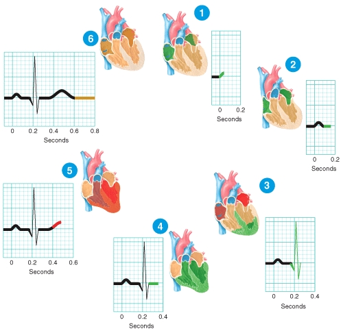
Briefly describe what is happening at the stage of the ECG labeled 5 in the diagram.
Free
(Essay)
4.9/5  (38)
(38)
Correct Answer: Verified
The contractile fibers of the ventricles are repolarizing,which generates the T wave in the ECG.
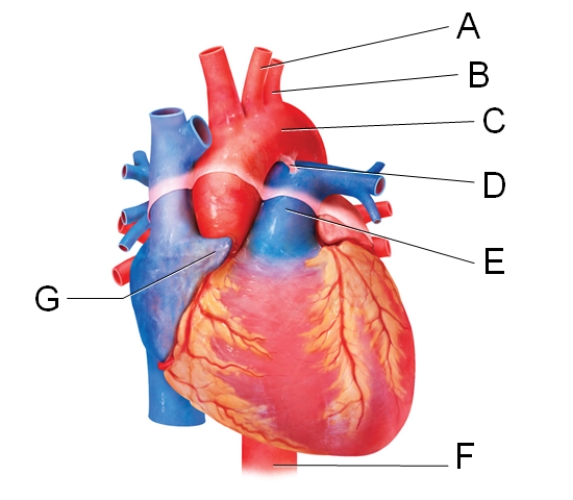
In the diagram,where is the left auricle of the left atrium?
Free
(Multiple Choice)
4.8/5 (33)
Correct Answer:Verified
C
During which of the following periods does the largest volume of blood enter the arteries?
Free
(Multiple Choice)
4.8/5 (25)
Correct Answer:Verified
D
The visceral layer of the serous pericardium is also considered to be the
(Multiple Choice)
5.0/5 (50)
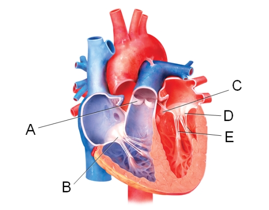
Which valve below prevents blood from flowing back into the right ventricle?
(Multiple Choice)
4.8/5 (32)
Contraction of the ventricles of the heart leads to blood moving directly
(Multiple Choice)
4.9/5 (37)
Isovolumetric contraction is the phase of the cardiac cycle in which
(Multiple Choice)
4.9/5 (34)
Which of the following is used to reduce friction between the layers of membranes surrounding the heart?
(Multiple Choice)
5.0/5 (37)
The membrane that surrounds and protects the heart is called the
(Multiple Choice)
4.8/5 (35)
Which labeled structure shown in the diagram is a remnant of fetal circulation that is not directly involved in adult circulation?

(Multiple Choice)
4.9/5 (42)
In comparison to skeletal muscle fibers,the contractile fibers of the heart are depolarized for [___] period of time.
Dropdown Choices:
a longer
a shorter
a variable
the same
(Short Answer)
4.7/5 (33)
The second heart sound (dupp)closely follows which of the events listed below?
(Multiple Choice)
4.8/5 (31)
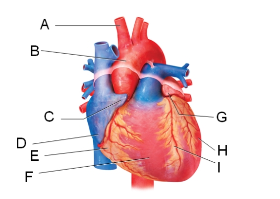
Which labeled structure in the figure is the ligamentum arteriosum? \

(Multiple Choice)
5.0/5 (40)
Which labeled structure shown in the diagram is a pouch-like extension that serves to slightly increase the capacity of an atrium?

(Multiple Choice)
4.8/5 (28)
The volume of blood ejected from the left ventricle into the aorta each minute is called the
(Multiple Choice)
4.8/5 (39)
Through which structure does blood pass from the right atrium to the right ventricle?
(Multiple Choice)
4.9/5 (22)
What is the function of the foramen ovale during fetal life?
(Multiple Choice)
4.8/5 (38)
Discuss the common nutrient sources used by cardiac muscle to produce ATP in a resting individual.
(Essay)
4.8/5 (34)
In the diagram,which labeled structure is the pulmonary semilunar valve?
(Multiple Choice)
4.8/5 (43)
Which labeled blood vessel shown in the diagram is the left common carotid artery?
(Multiple Choice)
4.9/5 (33)
Filters
- Essay(0)
- Multiple Choice(0)
- Short Answer(0)
- True False(0)
- Matching(0)