Exam 2: Observing the Microbial Cell
Exam 1: Microbial Life: Origin and Discovery70 Questions
Exam 2: Observing the Microbial Cell69 Questions
Exam 3: Cell Structure and Function72 Questions
Exam 4: Bacterial Culture, Growth, and Development70 Questions
Exam 5: Environmental Influences and Control of Microbial Growth70 Questions
Exam 6: Viruses70 Questions
Exam 7: Genomes and Chromosomes70 Questions
Exam 8: Transcription, Translation, and Bioinformatics76 Questions
Exam 9: Gene Transfer, Mutations, and Genome Evolution72 Questions
Exam 10: Molecular Regulation73 Questions
Exam 11: Viral Molecular Biology70 Questions
Exam 12: Biotechniques and Synthetic Biology72 Questions
Exam 13: Energetics and Catabolism77 Questions
Exam 14: Electron Flow in Organotrophy, Lithotrophy, and Phototrophy73 Questions
Exam 15: Biosynthesis73 Questions
Exam 16: Food and Industrial Microbiology73 Questions
Exam 17: Origins and Evolution70 Questions
Exam 18: Bacterial Diversity71 Questions
Exam 19: Archaeal Diversity70 Questions
Exam 20: Eukaryotic Diversity69 Questions
Exam 21: Microbial Ecology70 Questions
Exam 22: Microbes in Global Elemental Cycles70 Questions
Exam 23: Human Microbiota and Innate Immunity70 Questions
Exam 24: The Adaptive Immune Response70 Questions
Exam 25: Microbial Pathogenesis70 Questions
Exam 26: Microbial Diseases69 Questions
Exam 27: Antimicrobial Therapy72 Questions
Exam 28: Clinical Microbiology and Epidemiology75 Questions
Select questions type
Microbes were detected long before the invention of the microscope. How could this be?
Free
(Essay)
4.9/5  (40)
(40)
Correct Answer: Verified
Detection is the ability to observe the presence of an object, such as when we detect a group of bacteria in a culture tube or growing on a surface such as a food product. Even though we can detect the group, we cannot resolve individual cells without the magnification afforded by microscopes.
Why do some bacteria appear purple after being Gram stained, while others appear pink?
Free
(Essay)
4.8/5 (36)
Correct Answer:Verified
Gram-negative cells have a few layers of peptidoglycan cell wall and an outer lipopolysaccharide membrane. Gram-positive organisms have several layers of peptidoglycan and no outer membrane. The multiple layers of peptidoglycan retain the crystal violet-iodine complex, so appear purple. Gram-negative cells do not retain the crystal violet because there are few layers of peptidoglycan and the outer membrane is disrupted by the decolorizer.
Fluorescence microscopy using labeled antibodies is referred to as
Free
(Multiple Choice)
4.8/5 (31)
Correct Answer:Verified
A
When two waves are out of phase by ________ wavelength, they produce destructive interference, canceling each other's amplitude and resulting in contrast in the image.
(Multiple Choice)
5.0/5 (38)
Cryo-electron microscopy (cryo-EM) is differentiated from transmission electron microscopy because it
(Multiple Choice)
4.7/5 (38)
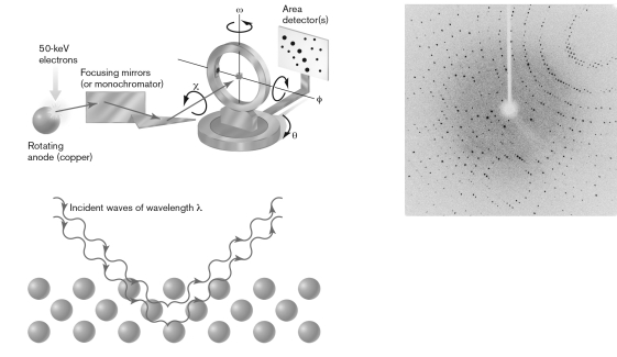
Using the figure below, explain how the visualization of molecules occurs through X-ray crystallography.
(Essay)
4.9/5 (42)
Define a fluorophore and give three examples of how it can be used to label cells.
(Essay)
4.8/5 (34)
If aqueous cytoplasm was submerged in a beaker of immersion oil, the slide would be
(Multiple Choice)
4.8/5 (29)
Which of these techniques can be used to localize the DNA sequence at the origin of replication in a bacterial cell?
(Multiple Choice)
4.9/5 (26)
Which of the following is best visualized using a negative stain?
(Multiple Choice)
4.8/5 (39)
Which of the following is an advantage of using chemical imaging microscopy?
(Multiple Choice)
4.8/5 (29)
What is the MOST important property that enables a lens to magnify an image?
(Multiple Choice)
4.7/5 (34)
Compare and contrast a simple stain (like methylene blue) with the Gram stain. What information about a microbial sample can be collected with each?
(Essay)
4.9/5 (35)
Fluorescent microscopy that absorbs light at 260m would MOST likely fluoresce at
(Multiple Choice)
4.9/5 (35)
Compare and contrast the radiation sources, lenses, and image-capturing devices used in light microscopy and transmission electron microscopy.
(Essay)
4.9/5 (36)
Which type of microscopy is used to identify the 3-D structure of biofilms?
(Multiple Choice)
4.9/5 (30)
Filters
- Essay(0)
- Multiple Choice(0)
- Short Answer(0)
- True False(0)
- Matching(0)