Exam 2: Functional Neuroanatomy: The Cells and Structures of the Nervous System
Summarize the neuron doctrine and the process of discovery that led up to it.
The neuron doctrine is a fundamental principle in neuroscience that states that the nervous system is composed of individual cells called neurons, which are the basic building blocks of the nervous system. This doctrine was developed through a process of discovery that involved several key milestones in the history of neuroscience.
One of the earliest discoveries that led to the neuron doctrine was made by the Spanish anatomist Santiago Ramón y Cajal in the late 19th century. Using a staining technique developed by Camillo Golgi, Cajal was able to visualize individual neurons and their connections in the brain. This led him to propose the idea that the nervous system is made up of discrete, separate cells rather than a continuous network of tissue.
Another important milestone in the development of the neuron doctrine was the work of the German anatomist Otto Deiters, who identified the structure of the neuron and its components, including the cell body, dendrites, and axon. This helped to further solidify the idea that neurons are distinct, individual cells.
The final piece of the puzzle came from the work of the British physiologist Charles Sherrington, who demonstrated that neurons communicate with each other through synapses, or specialized junctions where signals are transmitted from one neuron to another. This discovery provided further evidence for the idea that neurons are discrete entities that communicate with each other in a specific, organized manner.
Taken together, these discoveries and others led to the formulation of the neuron doctrine, which revolutionized our understanding of the nervous system and laid the foundation for modern neuroscience. The neuron doctrine has had a profound impact on our understanding of brain function and has been instrumental in shaping our current knowledge of the brain and nervous system.
Refer to the figure.
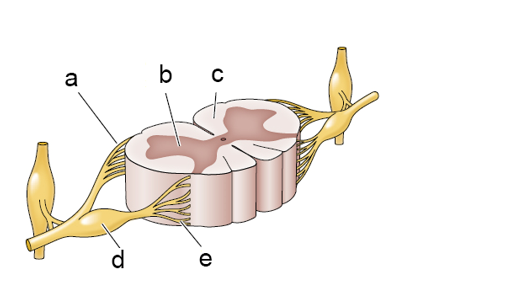 Match each letter label (a-e) on the spinal cord section to the anatomical label (1-5).
-_____
1. Dorsal roots (sensory)
2. Dorsal root ganglion
3. Ventral roots (motor)
4. Gray matter
5. White matter
Match each letter label (a-e) on the spinal cord section to the anatomical label (1-5).
-_____
1. Dorsal roots (sensory)
2. Dorsal root ganglion
3. Ventral roots (motor)
4. Gray matter
5. White matter
4
Which statement does not describe a reason that tract-tracing studies have been difficult to perform?
Although nerve cells typically have only one axon, an axon may divide into numerous axon
Located within the basal ganglia are the globus pallidus, caudate nucleus, and _______.
Describe the components and general function of the basal ganglia, limbic system, and reticular formation.
Although nerve cells typically have only one axon, an axon may divide into numerous axon _______.
The fornix and _______ are two components of the limbic system that form arcs under the surface of the cerebral hemispheres.
Compare and contrast at least four different human brain imaging technologies through discussion of their technical bases and the types of information each provides.
Axon terminals form the _______ side of a synapse, and dendrites form the _______ side of a synapse.
Filters
- Essay(0)
- Multiple Choice(0)
- Short Answer(0)
- True False(0)
- Matching(0)