Exam 6: Antigen Presentation to T Lymphocytes
The invariant chain protein, Ii, has only one function in MHC class II antigen presentation. This function entails Ii protein occupying the peptide-binding site of each newly synthesized class II protein, thereby preventing nascent MHC class II proteins from binding peptides or misfolded proteins in the endoplasmic reticulum.
False
NKT cells that recognize microbial glycolipids bound to CD1 molecules comprise a class of T cells that shares features of both innate and adaptive immune cells. A second class of such cells are MAIT cells, that recognize antigens bound to the MHC class Ib molecule, MR1. What is the class of PAMP recognized by MAIT cells?
Microbial products specific to folate metabolism.
Analysis of MR1 proteins that were refolded in the presence of supernatants from cultures of Salmonella typhimurium eventually led to the identification of several riboflavin metabolites that are formed by biosynthetic pathways in most bacteria and yeast. These metabolites not only bind to MR1, but also activate MAIT cells. Thus, MAIT cells are activated in response to infection by these organisms by detecting products specific to their folate metabolism. As such, MAIT cells appear to hold an intermediate place in the spectrum of innate and adaptive immunity, similar to iNKT cells, in that they use an antigen receptor assembled by somatic gene rearrangement, but recognize a molecular structure that falls within the definition of a PAMP.
During MHC class I synthesis and folding in the endoplasmic reticulum (ER), a process of peptide editing takes place as the newly synthesized MHC class I protein is held in a 'peptide receptive' state by binding to the calreticulin:ERp57:tapasin complex. Peptide editing ensures that the MHC class I molecules that reach the cell surface have stable, high affinity binding for their peptide cargo. Peptide editing is important to the immune response because it:
E
The experiment shown in Figure uses two strains of mice that differ in their MHC genes. Strain A is H-2a and Strain B is H-2b. Mice of each strain are infected with the virus LCMV, and T cells are isolated at day 8 post-infection. These T cells are mixed with target cells that express either H-2a or H-2b; in each case, the target cells are either uninfected or infected with LCMV. After a four-hour incubation of T cells with target cells, the percentage of target cells lysed by the T cells is shown in the graph. 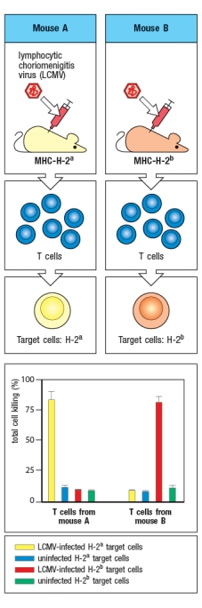 The explanation for the results of this experiment is:
Virus infections induce production of interferons that act on infected cells to enhance their recognition by CD8 cytotoxic T cells. To counter these mechanisms, viruses often encode proteins that interfere with antigen processing and presentation. In an experiment, cells infected with Virus X are treated with interferon and compared with uninfected cells treated with interferon. Proteasomes are isolated from the two cell populations and their enzymatic activities are compared. The data in Figure show the amino acid preferences for cleavage of peptides by the two samples of proteasomes. 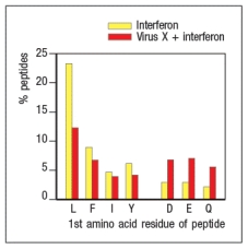 Based on these data, Virus X most likely encodes a protein that interferes with:
T cells expressing : T-cell receptors have been found to recognize a diversity of ligands, including pathogen-derived proteins, self-peptides, and stress-induced molecules. This pattern of antigen recognition shows similarity to that of iNKT cells and MAIT cells, suggesting that : T cells:
Empty MHC class I and MHC class II molecules are rapidly removed from the cell surface. This process prevents:
The adaptive immune system developed a strategy for monitoring the proteins synthesized in virtually any cell in the body, thereby preventing pathogens from 'hiding out' by adopting an intracellular lifestyle. To accomplish this, the immune system:
In a mixed lymphocyte reaction, T cells from individual A make a robust response to antigen-presenting-cells from individual B, as long as the two individuals express different alleles of MHC molecules. Estimates indicate that up to 10% of the T cells from individual A may contribute to this response. If one performed this assay using responder T cells from a child and antigen-presenting cells from one parent, the result would be:
MHC class I surface expression is dependent on an abundant source of pathogen-derived peptides. Thus, in uninfected cells, nearly all of the MHC class I proteins are degraded and never reach the cell surface.
A cell line carrying a mutation in a single gene is found to express very low levels of MHC class I on its surface. When infected with influenza virus, these cells are not recognized nor are they killed by a CD8 T cell line specific for an influenza peptide bound to the MHC class I protein expressed by these cells. Incubation of the mutant cell line with a large excess of this peptide in the cell culture medium overnight leads to the results shown in 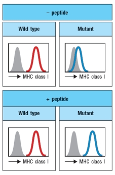 What is the most likely candidate for the gene that is defective in the mutant cell line?
Some viruses have mechanisms to down-regulate MHC class I protein expression on the surface of cells in which the virus is replicating. This immune evasion strategy might prevent effector CD8 cytotoxic T cells from recognizing and killing the virus-infected cells. Would this immune evasion strategy also prevent the initial activation of virus-specific CD8 T cells?
Some CD1 molecules bind to glycosphingolipids, and are recognized by a subset of T cells known as invariant NKT (iNKT) cells. The ability of these T cells to recognize different glycolipid constituents from microorganisms when they are bound to CD1d places these cells in the 'innate immune' category. While iNKT cells do express a fully rearranged : T-cell receptor, one key feature of the T-cell receptors expressed on iNKT cells also places them in the 'innate immune' category. This feature is:
NKG2D is an activating receptor expressed on NK cells, : T cells, and some cytotoxic : T cells. When stressed or infected cells up-regulate receptors that bind to and activate NKG2D molecules, the stressed or infected cells will be killed. This pathway relies on the fact that stressed or infected cells up-regulate:
The virus shown in the diagram below is only able to infect and replicate in epithelial cells. In order for the cross-presenting dendritic cell to display viral peptides, rather than self peptides on its surface MHC class I proteins, which of the following procedures could be utilized, starting with the components shown in Figure? 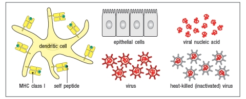
Peptide editing is an important component of antigen presentation for both MHC class I and MHC class II pathways, as it drives the preferential presentation of high-affinity binding peptides. For MHC class II peptide editing, HLA-DM plays a key role. In the absence of HLA-DM:
A family of six (mother, father, and four children) had two children with a history of chronic illness. Both children had repetitive infections of the sinuses, middle ears, and lungs due to a variety of respiratory viruses. Their other siblings were generally healthy and showed no signs of persistent or recurrent virus infections.
The two affected children had normal numbers of B cells, T cells, and NK cells in their blood. They also showed no defects in neutrophil function or in complement protein levels. The two children also had normal antibody levels to vaccine protein antigens, such as tetanus toxoid, and had normal T cell responses to antigens from the vaccine strain of Mycobacterium tuberculosis after being vaccinated.
Blood cells from one parent, one healthy child and the two affected children were examined for surface MHC protein expression by flow cytometry using two antibodies, one that recognizes all HLA class I proteins, and one that recognizes all HLA class II proteins. The results are shown in Figure
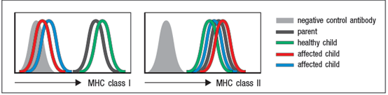 a) Analysis of HLA genotypes from the two affected children showed that they shared one haplotype of this locus. This haplotype encodes a common HLA-A allele, HLA-A2. Based on these data, is it likely that the two affected children have a point mutation (or mutations) in the coding sequence for HLA-A2? Why or why not?
b) Name two proteins that could be candidates for the defective gene in the two affected children.
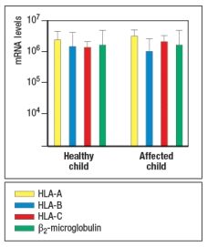
To address which gene defect might be present in the affected children, peripheral blood cells were isolated from one healthy child and one affected child, and mRNA was isolated from the cells. Quantitative RT-PCR was performed to assess the levels of mRNA for the three HLA class I heavy chain genes (HLA-A, -B, and –C) and for the β2-microglobulin gene. The results are shown in Figure .
a) Analysis of HLA genotypes from the two affected children showed that they shared one haplotype of this locus. This haplotype encodes a common HLA-A allele, HLA-A2. Based on these data, is it likely that the two affected children have a point mutation (or mutations) in the coding sequence for HLA-A2? Why or why not?
b) Name two proteins that could be candidates for the defective gene in the two affected children.
To address which gene defect might be present in the affected children, peripheral blood cells were isolated from one healthy child and one affected child, and mRNA was isolated from the cells. Quantitative RT-PCR was performed to assess the levels of mRNA for the three HLA class I heavy chain genes (HLA-A, -B, and –C) and for the β2-microglobulin gene. The results are shown in Figure .
 In a second experiment, Western blots were performed, confirming that cell lysates from both affected children contained normal levels of all three HLA class I heavy chain proteins and the β2-microglobulin protein.
c) Do these data eliminate any of your answers to part (b)?
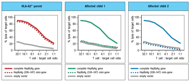
In a final experiment, peripheral blood cells from the two affected children and one HLA-A2+ parent were transfected with a construct encoding the Hepatitis B virus surface antigen (HepBsAg), a protein that is currently used in the vaccine against Hepatitis B. This protein is normally synthesized and transported to the cell surface. In addition to the full length HepBsAg construct, cells were also transfected with a mini-gene, encoding just a single HLA-A2-binding peptide derived from Hepatitis B, amino acids 338–347. Using a cytolytic CD8 T cell clone specific for HepBsAg(338–347) peptide bound to HLA-A2, the transfected cells were tested for recognition by the CD8 T cell clone using an assay that measures target cell killing. Figure Q6.29C shows the results of this experiment.
In a second experiment, Western blots were performed, confirming that cell lysates from both affected children contained normal levels of all three HLA class I heavy chain proteins and the β2-microglobulin protein.
c) Do these data eliminate any of your answers to part (b)?
In a final experiment, peripheral blood cells from the two affected children and one HLA-A2+ parent were transfected with a construct encoding the Hepatitis B virus surface antigen (HepBsAg), a protein that is currently used in the vaccine against Hepatitis B. This protein is normally synthesized and transported to the cell surface. In addition to the full length HepBsAg construct, cells were also transfected with a mini-gene, encoding just a single HLA-A2-binding peptide derived from Hepatitis B, amino acids 338–347. Using a cytolytic CD8 T cell clone specific for HepBsAg(338–347) peptide bound to HLA-A2, the transfected cells were tested for recognition by the CD8 T cell clone using an assay that measures target cell killing. Figure Q6.29C shows the results of this experiment.
 d) What is the most likely gene that is defective in the affected children?
The genes encoding MHC proteins are closely linked with genes encoding proteins involved in antigen processing and presentation. This genetic linkage facilitates the coordinate regulation of these genes by interferons.
The mechanism of cross-presentation by dendritic cells is an essential pathway for generating CD8 T cell responses to some intracellular pathogens. If this pathway did not exist, we would be highly susceptible to:
Three major cell types, dendritic cells, macrophages, and B cells, present peptides bound to MHC class II molecules for recognition by CD4 T cells. In general, these peptides are derived from proteins or pathogens taken up by the cell by endocytosis, phagocytosis, or macropinocytosis. Based on these pathways of antigen uptake, some of the enzymes that degrade proteins to generate peptides for MHC class II presentation are:
Filters
- Essay(0)
- Multiple Choice(0)
- Short Answer(0)
- True False(0)
- Matching(0)