Exam 5: The Generation of Lymphocyte Antigen Receptors
Exam 1: Basic Concepts in Immunology44 Questions
Exam 2: Innate Immunity: the First Lines of Defense32 Questions
Exam 3: The Induced Responses of Innate Immunity39 Questions
Exam 4: Antigen Recognition by B-Cell and T-Cell Receptors28 Questions
Exam 5: The Generation of Lymphocyte Antigen Receptors33 Questions
Exam 6: Antigen Presentation to T Lymphocytes30 Questions
Exam 7: Lymphocyte Receptor Signaling42 Questions
Exam 8: Development and Survival of Lymphocytes37 Questions
Exam 9: T-Cell-Mediated Immunity37 Questions
Exam 10: The Humoral Immune Response30 Questions
Exam 11: Integrated Dynamics of Innate and Adaptive Immunity28 Questions
Exam 12: The Mucosal Immune System27 Questions
Exam 13: Failures of Host Defense Mechanisms43 Questions
Exam 14: Allergy and Allergic Diseases26 Questions
Exam 15: Autoimmunity and Transplantation31 Questions
Exam 16: Manipulation of the Immune Response34 Questions
Select questions type
Classical MHC molecules function as peptide-binding receptors that present these peptides to : T cells for recognition by : T-cell receptors. MHC molecules can be detected as far back in evolution as sharks, the same time at which T-cell receptors can be identified. Examination of shark MHC proteins indicates that these molecules likely function identically to human MHC molecules. This conclusion is based on:
Free
(Multiple Choice)
4.8/5  (34)
(34)
Correct Answer: Verified
A
Several invertebrate species, such as some insect and snail species, have mechanisms for increasing the diversity of immune recognition molecules that are expressed in an individual beyond the simple 'one gene encodes one protein' rule that applies to most genes in the genome. One of these mechanisms:
Free
(Multiple Choice)
4.7/5 (37)
Correct Answer:Verified
E
Some pathogenic microorganisms encode proteins, such as the Staphylococcus Protein A, that bind to immunoglobulin constant region domains with high affinity. These microbial proteins provide a benefit to the microorganism by:
Free
(Multiple Choice)
4.9/5 (33)
Correct Answer:Verified
A
In some vertebrates, such as rays and some shark species, immunoglobulin light chain genes consist of multiple units of already rearranged VJ-C genes, of which one is chosen for expression in a developing B cell. This strategy may have evolved in these organisms:
(Multiple Choice)
4.9/5 (34)
Antibody diversity is generated by multiple mechanisms, each of which contributes to the generation of antibodies with up to 1011 different amino acid sequences in their antigen-binding sites. Several of these mechanisms involve changes in the DNA sequences encoding the antibody heavy and light chain proteins. One mechanism that does not rely on changes to the DNA within the immunoglobulin heavy and light chain gene loci is, instead, dependent on:
(Multiple Choice)
4.9/5 (34)
IgM is the first antibody isotype secreted following activation of a naive B cell. IgM is found at high concentrations in the serum, and is found as a very high molecular weight complex. This high molecular weight complex is composed of:
(Multiple Choice)
4.9/5 (32)
While B cells and T cells differ markedly in their functions during an immune response, the two lymphocyte subsets share the enzymatic machinery and overall scheme for generating antigen receptor diversity. This is because:
(Multiple Choice)
4.7/5 (36)
Immunodeficiency diseases arise when individuals lack one or more components of their immune system, and are identified by an individual's history of persistent or recurrent infections. Some genetic defects (mutations or small deletions) can cause profound defects in an immune cell population; alternatively, in some cases, such small defects occur that there is no visible effect on immune responses. The diagram in Figure shows simplified versions of the immunoglobulin heavy chain locus, the T-cell receptor chain locus, and the locus encoding the RAG-1 and RAG-2 recombinases. For the sake of this question, imagine that these diagrams represent all of the gene segments present in the immunoglobulin heavy chain and T-cell receptor chain locus. 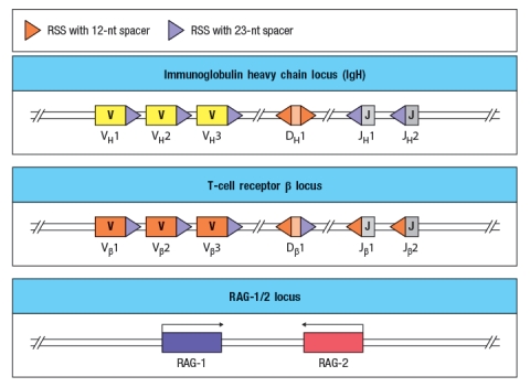 You now analyze five individuals, each of which has a single inactivating mutation in a region of one of these three loci. These mutations are each indicated by a red 'X' in Figure , and are numbered 1-5. For each of these inactivating mutations, indicate the alterations and/or defects that would be seen in the repertoire of antigen receptors found in mature B and T cells in that individual. Also, for each mutation, indicate whether the individual would likely show any immunodeficiency, such as a history of recurrent infections.
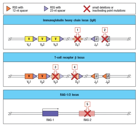
(Essay)
4.8/5 (33)
Antibodies that bind with high affinity to some viral surface proteins require heavy chain CDR3 loops of unusual length. Whereas the average human heavy chain CDR3 length is ~15 amino acids, antibodies with VH CDR3 loops of >30 amino acids are readily detected in the repertoire. These antibody heavy chains with CDR3 lengths of >30 amino acids would likely be missing in individuals lacking:
(Multiple Choice)
4.9/5 (43)
Most eukaryotic genes are encoded in a set of exons that are brought together to form a contiguous protein coding sequence by the process of mRNA splicing. In contrast, immunoglobulin genes use somatic recombination of gene segments and not mRNA splicing to generate the final mRNA that is translated into protein.
(True/False)
4.8/5 (36)
The exon encoding the V region of an immunoglobulin protein is generated by a process of somatic recombination. This recombination event brings V gene and J gene segments together:
(Multiple Choice)
4.8/5 (47)
The generation of a complete coding sequence for an antibody heavy chain involves a lymphocyte-restricted process of DNA rearrangement that links V, D, and J gene segments together to form the exon that encodes the heavy chain V region. A similar type of DNA rearrangement is also utilized for the simultaneous expression of IgM and IgD antibodies by the same B cell.
(True/False)
4.9/5 (33)
For immunoglobulin heavy and light chain genes, and for T-cell receptor chain genes, there are a large number of V gene segments, and relatively few J and/or D segments that rearrange to form the final coding sequence for each gene. The TCR locus is different in this regard, and this difference is thought to reflect the fact that nearly all : T-cell receptors recognize a peptide bound to an MHC molecule. This unique feature of the T-cell receptor locus is:
(Multiple Choice)
4.8/5 (31)
B-cell receptors and T-cell receptors share a mechanism for generating diversity, and also share overall structural homology both in their V domains and their C domains. This is because the two proteins have nearly identical functions in the immune responses mediated by their respective cell types.
(True/False)
4.9/5 (34)
When a B cell differentiates into a plasma cell, it goes from expressing membrane-bound IgM to making mostly the secreted form of IgM. A deletion of the first polyadenylation site in the μ heavy chain gene would prevent activated B cells from making the secreted form of IgM.
(True/False)
4.8/5 (39)
An important mechanism for generating diversity in immunoglobulin light chain V-region sequences is based on the fact that the RAG recombinase generates hairpin structures, rather than blunt ends, at the cleavage sites between the recombination signal sequences and the coding sequences. Explain how this mechanism generates diversity at the junctions.
(Essay)
4.8/5 (27)
The V-D-J recombination process used to generate antigen receptor diversity in vertebrates is believed to have evolved from a transposon. How does this hypothesis explain why the recombination signal sequences of the antigen receptor gene segments are joined precisely during the recombination process, whereas the cut ends that are joined to form the antigen receptor coding sequence are joined by an error-prone process?
(Essay)
4.8/5 (41)
The evolutionary conservation of two classes of T lymphocytes, those expressing : T-cell receptors and those expressing : T-cell receptors, argues for important but separate functions of these two classes of T cells. Evolution has also conserved the process of somatic recombination of gene segments that is able to generate an enormous diversity of : and : T-cell receptor sequences. Do these observations indicate that both classes of T cells are components of the adaptive immune system? Why or why not?
(Essay)
4.8/5 (35)
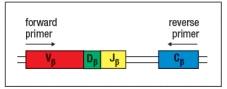
T-cell receptor spectratype analysis is used to examine the diversity of T-cell receptor chain sequences in an individual's T cells. For this technique, T cells are isolated from a sample of thymocytes (developing T cells) or mature peripheral T cells from an individual. The mRNA is isolated from these cells and cDNA is generated by reverse transcription. This pool of cDNA is mixed with PCR primers that are used to amplify part of the rearranged T-cell receptor chain sequence containing the complete CDR3. The position of these primers relative to the rearranged T-cell receptor chain gene in the DNA locus is shown in Figure. Following the PCR amplification, the heterogeneous mixture of DNA molecules is then size-separated by electrophoresis on an apparatus that can separate molecules that differ by a single nucleotide. At the end, the quantity of material deposited in each band of a given nucleotide sequence length is quantified by densitometry, and the spectratype trace is produced. The x-axis of the spectratype depicts the number of nucleotides in each PCR product from the beginning of the forward primer to the end of the reverse primer.
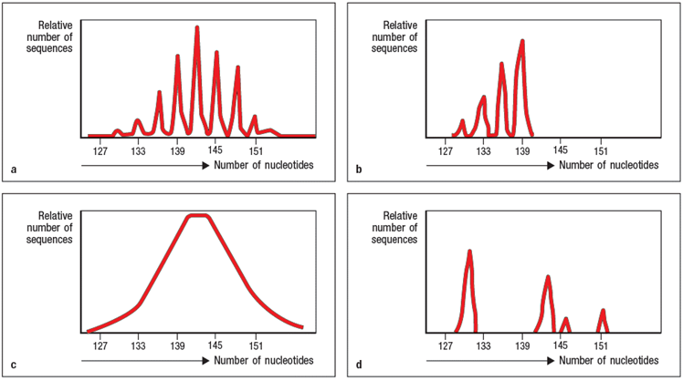 a) Panel A of Figure shows the spectratype trace of mature peripheral T cells from a healthy individual. What is explanation for the separation of the heterogeneous population of T-cell receptor chain sequences into multiple sharp peaks of size lengths?
b) Panel B shows T cells from an individual that is missing an important enzyme that contributes to T-cell receptor chain diversity during the recombination process. Which enzyme is most likely absent in this individual?
c) Panel C shows the spectratype analysis of T-cell receptor chain sequences in developing T cells that have just completed the V-D-J recombination process. Explain why this spectratype looks different from the one shown in panel A.
d) Panel D shows a more restricted example of spectratype analysis, where the forward primer used only binds to one specific V sequence. In this example, the primer is specific for V 17. When such a V -specific primer is used, the spectratype analysis only shows the junctional sequence lengths for T cells whose chain uses V 17. In a healthy individual, the V 17 spectratype would like just like the one shown in panel A; in other words, it would show a random distribution of V 17+ T-cell receptor chains with a normal distribution of junctional lengths.
However, in this case, the individual being studied has been infected with influenza virus, and is in the midst of a robust T cell response against the virus. What is the likely explanation for the non-random pattern of peaks on the V 17 spectratype from this individual at this timepoint?
a) Panel A of Figure shows the spectratype trace of mature peripheral T cells from a healthy individual. What is explanation for the separation of the heterogeneous population of T-cell receptor chain sequences into multiple sharp peaks of size lengths?
b) Panel B shows T cells from an individual that is missing an important enzyme that contributes to T-cell receptor chain diversity during the recombination process. Which enzyme is most likely absent in this individual?
c) Panel C shows the spectratype analysis of T-cell receptor chain sequences in developing T cells that have just completed the V-D-J recombination process. Explain why this spectratype looks different from the one shown in panel A.
d) Panel D shows a more restricted example of spectratype analysis, where the forward primer used only binds to one specific V sequence. In this example, the primer is specific for V 17. When such a V -specific primer is used, the spectratype analysis only shows the junctional sequence lengths for T cells whose chain uses V 17. In a healthy individual, the V 17 spectratype would like just like the one shown in panel A; in other words, it would show a random distribution of V 17+ T-cell receptor chains with a normal distribution of junctional lengths.
However, in this case, the individual being studied has been infected with influenza virus, and is in the midst of a robust T cell response against the virus. What is the likely explanation for the non-random pattern of peaks on the V 17 spectratype from this individual at this timepoint?
(Essay)
4.8/5 (44)
Different individuals can have different numbers of functional V gene segments as well as different numbers of constant region genes. This type of genetic polymorphism between individuals indicates that:
(Multiple Choice)
4.9/5 (32)
Filters
- Essay(0)
- Multiple Choice(0)
- Short Answer(0)
- True False(0)
- Matching(0)